The Oaks

The Oaks

Etiketter

måndag 3 april 2023

Aloe vera tutkimuksista

 https://link.springer.com/chapter/10.1007/978-3-0348-0927-6_6

Home

The Genus Aloe: Phytochemistry and Therapeutic Uses Including Treatments for Gastrointestinal Conditions and Chronic Inflammation

Part of the Progress in Drug Research book series (PDR,volume 70)

Abstract

Plants of the genus Aloe have perhaps the longest recorded history of medicinal usage and are amongst the most widely used plants for traditional medicinal purposes worldwide. Aloe vera , Aloe ferox , Aloe arborescens and Aloe perryi are the best known and most widely used, but many other species are also used for their therapeutic properties. The Aloes have been used since ancient times, particularly for the treatment of microbial infections, gastrointestinal disorders and inflammatory conditions. In addition to their myriad uses in traditional therapeutics, the Aloes have also been used as components of cosmetic formulations, and in the food and beverage industries. Despite their wide acceptance, studies from different laboratories often report wide variations in the therapeutic bioactivities from within the same Aloe species, even when the same extraction procedures are used. Furthermore, leaves from individual Aloe plants within the same species may have widely varying levels of the bioactive phytochemicals. Phytochemical analyses have shown that many Aloe species contain various carbohydrate polymers (notably glucomannans ) and a range of other low molecular weight phenolic compounds including alkaloids , anthraquinones , anthrones , benzene and furan derivatives , chromones , coumarins , flavonoids , phytosterols , pyrans and pyrones . There has been a wealth of information published about the phytochemistry and therapeutic potential of the Aloes (especially Aloe vera). Much of this has been contradictory. Intra- and interspecies differences in the redox state of the individual Aloe components and in the ratios of these components may occur between individual plants. These factors may all affect the physiological properties of Aloe extracts. Due to the structure and chemical nature of many of the Aloe phytochemicals, it is likely that many of the reported medicinal properties are due to antioxidant or prooxidant effects. The antioxidant/prooxidant activities of many Aloe phytochemicals depend not only on their individual levels, but also on the ratios between the various components and their individual redox states. Therefore, discrepancies between bioactivity studies are likely when using different crude mixtures. This report aims to summarise the phytochemistry of the Aloes and (a) examine how their constituents may be responsible for their medicinal properties and (b) some possible reasons for the wide variations reported for their medicinal properties and (c) their therapeutic mechanisms. Some future areas of research into the medicinal activities of this important genus are also highlighted.

Keywords

  • Aloe vera
  • Inflammation
  • Anticancer
  • Antioxidant
  • Anthraquinone
  • Anthrone

This is a preview of subscription content, access via your institution.


fredag 3 mars 2023

Eteerisen kuminaöljyn tuoksuinen karvoni, Carvone, Karvon, on terpeeniketoni

 http://www.shenet.se/ravaror/karvon.html

Doftbeskrivning
1) D-karvon: Luktar eterisk kumminolja.
2) L-karvon: Luktar eterisk grönmyntaolja men sötare och renare.
Användning
1) D-karvon: Kummindofter använda mycket i mat; kan ingå med upp till 2 % av doftämnena.
2) L-karvon: Använda i friska dofter där den kan utgöra upp till 3 % av doftämnena.

insektsmedel: Stöter bort insekter.

Kramplösande på glatt muskulatur, vilket är en del av förklaringen till att eteriska oljor som kumminessens är till hjälp vid kolik.

Mat och dryck
Smakämnen i matindustrin - ger kummin- respektive myntasmak.
Giftighet
Som så många ketoner - ja.
1) d-karvon: FAO/WHO har satt gränsen för acceptabelt dagligt intag vid 1 mg per kilo kroppsvikt. Akut giftighet prövades i en studie i början av 80-talet; 800 mg olja per kilo kroppsvikt injicerad på möss framkallade kramper. Översatt till människa (70 kilo): injicerar man 54 gram karvon (56 ml) får man troligen kramper.
2) l-karvon: Åtminstone en av l-karvonerna uppges vara giftig, men inte mer än att den används som smakämne i livsmedel. 


Farmakopéerna
Karvon ersatte eterisk kumminolja i 1901 och 1908 års upplagor av svenska farmakopén men kumminoljan togs tillbaka 1925. I Tyskland där karvon var officinellt kring förra sekelskiftet kallades det Oleum carvi, kumminoljans farmakopénamn i andra länder.
Framställning 1) D-Karvon
• Källor: Utgör den tyngre delen av kumminessens (50-60 %), mer ju mognare fröna är. Destilleras också ur dillfröessens (35-60 %).

• Tillverkning: Terpenketon (monoterpen och omättad keton) som oxideras (syre upptas, väte avges) ur terpenen L-limonen (vänstervridande limonen).
2) L-Karvon
• Källor: Mest i grönmyntaessens (50-70 %) varur det extraheras.
• Tillverkning: Terpenketon (monoterpen och omättad keton) som oxideras ur terpenen D-limonen (högervridande limonen).
Beskrivning
Båda är färglösa till ljusgula vätskor som gulnar i kontakt med luft. De är kemiskt identiska men spegelvända och luktar olika.

100 ml väger ca 96 gram. 100 gram = ca 104 ml.

• Lösliga i 48-50 % alkohol (löses klart i 20 delar), vegetabilisk olja, mineralolja, propylenglykol.
• Olösliga i vatten, glycerin.
Innehåll Om ovanstående alkohollösning grumlas är karvonet förorenat av karven, den rest som återstår i kumminolja när karvon tagits bort; bestående huvudsakligen av limonen.
Varianter
• Många ytterligare former - dl-, pino-, iso-, cis-, trans-...
Ersättning
Kumminolja kan ersätta och ersättas av karvon. T. ex. tillät 1901 och 1908 års svenska farmakopéer att karvon utlämnades om recept angav kumminolja.
Hållbarhet Förvaras som eteriska oljor - mörkt, svalt, tillslutet. Inte särskilt hållbart. Gul färg är ett tecken på att karvonet är gammalt eller har förvarats fel. D-karvon används för att hindra potatis från att gro och mögla under lagring.
Inköp We don't sell, we tell!
Inget man hittar i svenska butiker.


fredag 10 juli 2020

RhoGTPaasi, Geranylgeranyltransferaasi-1, podosyytti , Bpix

https://gupea.ub.gu.se/handle/2077/64541

A Podocyte view on RhoGTPases and actin cytoskeleton regulation

  Amplification of the Melanocortin-1 Receptor In Nephrotic Syndrome Identifies a Target for Podocyte Cytoskeleton Stabilization. Bergwall L, Wallentin H, Elvin J, Liu P, Boi R, Sihlbom C, Hayes K, Wright D, Haraldsson B, Nyström J and Buvall L. Scientific Reports (2018) 8 (1), 15731
VISA ARTIKEL


II: Podocyte Geranylgeranyl transferase type I is essential for maintenance of the glomerular filtration barrier function. Bergwall L, Boi R, Akula M.K, Ebefors K, Bergo O.M, Nyström J, Buvall L. Manuscript.

The Role of Beta-pix in podocyte Rac1 activation and cytoskeleton rearrangement. Bergwall L, Wallentin H, Boi R, Svensk S, Lövljung V, Sihlbom C, Weins A, Ericsson A, William-Olsson L, Granqvist B. A, Ebefors K, Nyström J, Buvall L. Manuscript.

Fredagen den 4 september 2020, kl 9.00, Hjärtats Aula, Sahlgrenska Universitetssjukhuset, Göteborg
Proteinuria is a hallmark symptom of chronic kidney disease, that if left to persist constitutes a risk for progression of disease. Symptomatic treatment aiming at decreasing proteinuria is therefore standard practice. Curative treatments for the underlying cause of disease are however lacking and treatments currently in use to induce disease remission are associated with unfavorable side effects. Dysregulation of the podocyte actin cytoskeleton underlies the pathological process called foot process effacement (FPE), which is one of the leading causes of proteinuria. The studies included in this thesis have focused on podocyte actin cytoskeleton regulation and a group of proteins called RhoGTPases, known to be involved in actin cytoskeleton regulation in podocytes. In the first study, glomerular microarray analysis showed an increase in the expression of the melanocortin 1-receptor (MC1R) in renal diseases focal segmental glomerulosclerosis and membranous nephropathy. Subsequent mass spectrometry analysis in combination with pathway and biochemical analysis revealed the podocyte protective effects of MC1R stimulation in vitro. Activation of MC1R proved to be stabilizing the podocyte actin cytoskeleton through inhibition of the epidermal growth factor receptor (EGFR) and maintenance of the actin associated protein synaptopodin. In the second study, the depletion of the prenylation enzyme Geranylgeranyl transferase type I (GGTase-I) in podocytes led to the development of proteinuria and FPE in mice due to an imbalanced RhoGTPase activity and disruption of the actin cytoskeleton. These findings suggest that GGTase-I activity is essential for podocyte function. In the last study, a guanine nucleotide exchange factor (activator of RhoGTPases) named Bpix was identified to be modulated in podocytes following treatment with a renal stressor, using mass spectrometry analysis. Gene silencing of Bpix protected against actin cytoskeleton remodulation in a model of podocyte injury, demonstrating the importance of Bpix for podocyte actin cytoskeleton regulation. In conclusion, the results in this thesis confirm the importance of actin cytoskeleton regulation for podocyte integrity. Further on, the results provide new information on actin cytoskeleton regulatory pathways involving RhoGTPases in podocytes, which can be of importance for future attempts in finding targeted treatments of proteinuria and chronic kidney disease.

torsdag 2 juli 2020

Prenylaatio on posttranslationaalinen modifikaatio eräisiin proteiiniin.

Otan sitaattina vielä netistä biologian opetusta prenylaatiosta, sillä  SARS2-viruksella on muutamia interaktioproteiineja  prenyloiduissa ihmisen proteiineissa kuten    RAB14 ( Ras onkogeeni-perheen jäsen) , RALA( Ras like proto-onkogeeni A), RAB5C, RAB7A, RAB2A, RAB10, RHOA (Ras homologisen  perheen jäsen A).
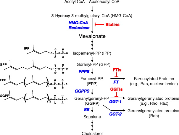
Prenylaatiotie taas on  maailmassa yleisesti  lääkkein vaikutettuna, koska sen tien päädyssä on koplesterolisynteesi, jota koetetaan säätää.  Mitä lääkesäätö vaikuttaa   prenylaation normaalikarttaan kehossa, on toinen asia. Prenylaatiotie on  kompromittoitunut syövissä.
https://media.springernature.com/lw685/springer-static/image/art%3A10.1007%2Fs12035-013-8627-z/MediaObjects/12035_2013_8627_Fig1_HTML.gif

https://what-when-how.com/molecular-biology/prenylation-molecular-biology/

Prenylation (Molecular Biology)

Prenylation or isoprenylation is a post-translational modification (PTM) process in which cysteine residues close to the C-terminal regions of some eukaryotic proteins are 
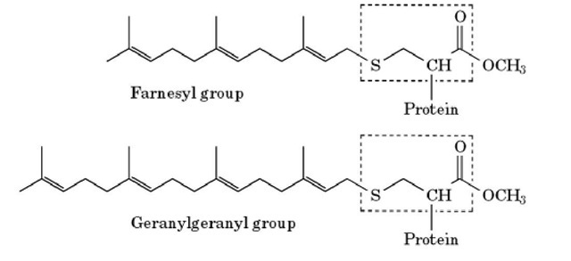
biosynthetically modified with an isoprenoid lipid: the 15-carbon farnesyl group or the 20-carbon geranylgeranyl group (see Fig. 1 and Table 1). Prenylation provides some proteins with a hydrophobic membrane anchor, and is important for their correct localization within the cell. Prenylation is one of several processes that attach lipid membrane anchors to proteins (see Membrane Anchors).

Figure 1. Modification of C-terminal cysteine residues by prenyl groups. The C-terminal cysteine residue of the protein is outlined by the dotted line. The thiol group is thioether-linked to either a farnesyl or a geranylgeranyl group, and the exposed carboxyl group is methylated.
 
http://what-when-how.com/wp-content/uploads/2011/05/tmp1C12_thumb.jpg 


Table 1. Examples of Prenylated Proteins
 
Farnesylated
Ras proteins
Transducin g subunit
Rhodopsin kinase
Nuclear lamins A and B
Fungal mating pheromonesa
Geranylgeranylated
g subunits of heterotrimeric G-proteins
Ras-related G-proteins (Rho/Rac/Rap/Ral/Rab)

Isoprenoids are branched unsaturated hydrocarbons that are synthesized in eukaryotic cells from acetyl Coenzyme A (Acetyl CoA)  by the first part of the metabolic pathway that is used to synthesize cholesterol and other sterols.
Attachment of isoprenoids to proteins is a post-translational process with four main steps:
 1) recognition of the C-terminal sequence (CAAX)  by one of three distinct prenyltransferases (1);
 2) prenylation of a cysteine (C)  residue(s) located at or close to the C-terminus using farnesylpyrophosphate  (FPP) or geranylgeranylpyrophosphate (GGPP)  as the substrate;
 3) proteolysis of the C-terminal residues  (-AAX) exposes the carboxyl group on the prenylated cysteine; and
 4) the isoprenylated cysteine is recognized by a methyltransferase, which methylates the carboxyl group using S-adenosyl methionine (SAM) as the methyl donor.
Steps 1) to 3) take place in the cytosol, whereas step 4) occurs on the cytoplasmic surface of the endoplasmic reticulum (ER) or the plasma membrane (PM) .
 Thus efficient methylation requires prior isoprenylation to localize the protein at the membrane surface. The thioether linkage between the cysteine and the prenyl group is chemically very stable and probably not subject to metabolic turnover. However, the carboxylic ester linkage to the methyl group is relatively labile, and may be removed after attachment. These steps differ substantially between proteins, depending on the sequence motif at the C-terminus:
 
1. Cys-a-a-X (CAAX)  If X is serine (S) , methionine (M), or glutamine (E), it is recognized by farnesyl transferase (FTase), and the cysteine residue will be farnesylated.
If X is leucine (L), it is recognized by geranylgeranyltransferase I (GGTase-1), and the cysteine residue will be geranylgeranylated. The identity of the "a" residues (usually aliphatic) is less important, but can influence whether isoprenylation takes place or not.
Farnesyl transferase and geranylgeranyltransferase I are both heterodimers; they have identical a subunits, whereas the a subunits have only 30% identify.
Farnesylation can also occur at the C-terminus of a variety of fungal mating pheromone peptides, and in yeast the same enzyme is used for farnesylating both proteins and peptides. Although farnesyl groups have relatively low affinity for membranes themselves, they can enhance the membrane association due to other lipid groups. Farnesyl groups, because of their small size, may also play an important role in protein-protein interactions by binding directly to specific sites on other proteins (2, 3).


2. Cys-Cys, Cys-X-Cys or Cys-Cys-X-X.
(CC, CXC or CCXX)  These double cysteine motifs (CC)  are restricted to the Rab subgroup of Ras-related small G-proteins. The Rab protein first forms a complex with Rab escort protein (REP1, CHM).
The Rab-REP complex is then recognized by geranylgeranyltransferase II.
 After prenylation, REP remains bound to Rab until it is delivered to the membrane.
 REP (CHM)  probably has a dual role: recognition of Rab and masking the two geranylgeranyl groups until they can be inserted into the appropriate membrane. Both cysteines are geranylgeranylated, and consequently proteolysis cannot occur. The C-terminus is not methylated in those Rab proteins ending with the sequence Cys-Cys (4).

Many of the prenylated proteins are involved in signal transduction or vesicle traffic, and the prenyl group, by facilitating rapid and reversible binding to membranes, plays an essential role in these functions (5, 6).
The membrane affinity of the prenylated proteins can be influenced by four different mechanisms (for a general discussion of factors which can affect membrane affinity of lipid anchored proteins, see Membrane Anchors):
1. The attachment of a palmitate residue (see Palmitoylation) to a cysteine close to the C-terminus reinforces the binding (eg, as in H- or N-Ras). Palmitoylation only occurs in membranes, however, so prenylation is required for it to take place (7).
2. The presence of basic residues close to the C-terminus will result in electrostatic attraction to the negatively charged bilayer surface (as in K-Ras) and increase membrane affinity (8).
3. Methylation converts the C-terminal residue from a negatively charged, hydrophilic group to an uncharged, hydrophobic group and increases membrane affinity approximately 10-fold (5, 6)). The increase in affinity is due to the hydrophobicity of the methyl group, rather than a reduction in electrostatic repulsion, because methylation gives comparable increases in binding to uncharged membranes. Methylation can have a profound influence on the cellular distribution of farnesylated proteins, because the farnesyl group is too short to provide an effective anchor by itself. Turnover of the methyl group has also been observed, and it is possible that repeated cycles of methylation and demethylation are used to regulate protein function.
4. The membrane affinity will be reduced by soluble carrier proteins, which are able to bind to the isoprenyl group(s) and mask them from the aqueous environment. This mechanism is important for the repeated releasing and recycling of Rab proteins during membrane vesicular traffic processes (9, 10)).
Previous post:

Zolendronaatti vaikuttaa prenylaatioon ja mm. sitä tietä voi olla eduksi covid-19 hoidossa

https://pubmed.ncbi.nlm.nih.gov/32600410/
Review
doi: 10.1186/s12967-020-02433-6.
 Boning Up: Amino-Bisphophonates as Immunostimulants and Endosomal Disruptors of Dendritic Cell in SARS-CoV-2 Infection
Affiliations
PMID: 32600410
PMCID: PMC7322393
DOI: 10.1186/s12967-020-02433-6
Free PMC article
 Abstract
Amino-bisphosphonates such as zoledronic acid (ZA) can possibly ameliorate or prevent severe COVID-19 disease by at least three distinct mechanisms: (1) as immunostimulants which could boost γδ T cell expansion, important in the acute response in the lung; (2) as DC modulators, limiting their ability to only partially activate T cells; and (3) as prenylation inhibitors of small GTPases in the endosomal pathway of the DC to prevent expulsion of lysosomes containing SARS-CoV-2 virions. Use of ZA or other amino-bisphosphonates as modulators of COVID-19 disease should be considered.
Keywords: Bisphosphonates; Glycoproteins; Immune response; SARS Coronavirus.
 Conflict of interest statement
AB is a paid consultant for Novartis, Amgen, and Sandoz. MTL, JG, and AR have no competing interests.
Zoledronic acid (ZA) acts as immunostimulant and endosomal disruptor of dendritic cell in SARS-CoV-2 infection. Inhaled SARS-CoV-2 particles are internalized by the DC (top). In COVID-19 disease, there is depletion of γδ T cells (bottom-left). In addition, virion release depends on prenylation signaling derived from the mevalonate pathway. On the other hand, ZA (bottom-right) inhibits the conversion of geranyl pyrophosphate (GPP) to farnesyl pyrophosphate (FPP), increasing the concentrations of isopentenyl pyrophosphate (IPP). Release of IPP induces γ9δ2 T-cell expansion by phosphoantigen recognition, mediated by butyrophilin-presentation. Downstream inhibition of prenylation reduces the activity of GTPases, decreasing the release of SARS-CoV-2. ZA also affects differentiation of the DC with downregulation of the expression of CD1a, CD11c, CD83, CD86, DC-SIGN, and HLA-DR and enhancement of the expression of CD80. Figure was created using BioRender https://biorender.com/

RAB perheen, Ras onkogeenin kaltaisen perheen prenylaatiosta

1)    CHM, (Xq21.2) , Rab proteins geranylgeranyltransferase component A1, (REP-1)

https://www.genecards.org/cgi-bin/carddisp.pl?gene=CHM&keywords=Rab,prenylation
Aliases for CHM Gene
CHM Rab Escort Protein 2 3 5
Rab Proteins Geranylgeranyltransferase Component A 1 3 4
Choroideremia (Rab Escort Protein 1) 2 3
CHM, Rab Escort Protein 1 2 3
Rab Escort Protein 1 2 4
REP-1 3 4
TCD 3 4
Choroideremia Protein 4
TCD Protein 4
HSD-32 3
DXS540 3
GGTA 3
REP1 4


Entrez Gene Summary for CHM Gene
This gene encodes component A of the RAB geranylgeranyl transferase holoenzyme. In the dimeric holoenzyme, this subunit binds unprenylated Rab GTPases and then presents them to the catalytic Rab GGTase subunit for the geranylgeranyl transfer reaction. Rab GTPases need to be geranylgeranyled on either one or two cysteine residues in their C-terminus to localize to the correct intracellular membrane. Mutations in this gene are a cause of choroideremia; also known as tapetochoroidal dystrophy (TCD). This X-linked disease is characterized by progressive dystrophy of the choroid, retinal pigment epithelium and retina. Alternatively spliced transcript variants have been found for this gene. [provided by RefSeq, Mar 2016]
GeneCards Summary for CHM Gene
CHM (CHM Rab Escort Protein) is a Protein Coding gene. Diseases associated with CHM include Choroideremia and Retinitis Pigmentosa. Among its related pathways are Metabolism of proteins and RAB GEFs exchange GTP for GDP on RABs. Gene Ontology (GO) annotations related to this gene include GTPase activator activity and Rab geranylgeranyltransferase activity. An important paralog of this gene is CHML.
UniProtKB/Swiss-Prot Summary for CHM Gene: Substrate-binding subunit of the Rab geranylgeranyltransferase (GGTase) complex. Binds unprenylated Rab proteins and presents the substrate peptide to the catalytic component B composed of RABGGTA and RABGGTB, and remains bound to it after the geranylgeranyl transfer reaction. The component A is thought to be regenerated by transferring its prenylated Rab back to the donor membrane. Besides, a pre-formed complex consisting of CHM and the Rab GGTase dimer (RGGT or component B) can bind to and prenylate Rab proteins; this alternative pathway is proposed to be the predominant pathway for Rab protein geranylgeranylation. RAE1_HUMAN,P24386

2) RABAC1,  RAB Acceptor 1
 https://www.genecards.org/cgi-bin/carddisp.pl?gene=RABAC1&keywords=Rab,prenylation
 Aliases for RABAC1 Gene
Rab Acceptor 1 2 3 5
Prenylated Rab Acceptor Protein 1 3 4
Rab Acceptor 1 (Prenylated) 2 3
Prenylated Rab Acceptor 1 2 3
PRA1 Family Protein 1 3 4
PRA1 Domain Family 1 2 3
PRAF1 3 4
PRA1 3 4
YIP3 3

GeneCards Summary for RABAC1 Gene
RABAC1 (Rab Acceptor 1) is a Protein Coding gene. Diseases associated with RABAC1 include Pontocerebellar Hypoplasia, Type 3 and Subvalvular Aortic Stenosis. Gene Ontology (GO) annotations related to this gene include identical protein binding and proline-rich region binding.
UniProtKB/Swiss-Prot Summary for RABAC1 Gene
General Rab protein regulator required for vesicle formation from the Golgi complex. May control vesicle docking and fusion by mediating the action of Rab GTPases to the SNARE complexes. In addition it inhibits the removal of Rab GTPases from the membrane by GDI. PRAF1_HUMAN,Q9UI14
  • (3)  Some RAB  Ras oncogene family membres: 
 RAB14 (9q33.2) (SARS2  nsp7  interaction protein)
Aliases for RAB14 Gene
RAB14, Member RAS Oncogene Family 2 3 5
BA165P4.3 (Member RAS Oncogene Family) 2 3
Small GTP Binding Protein RAB14 2 3
F Protein-Binding Protein 1 2 3
Ras-Related Protein Rab-14 3 4
RAB-14 3
FBP 3
RAB14 Gene: Involved in membrane trafficking between the Golgi complex and endosomes during early embryonic development. Regulates the Golgi to endosome transport of FGFR-containing vesicles during early development, a key process for developing basement membrane and epiblast and primitive endoderm lineages during early postimplantation development. May act by modulating the kinesin KIF16B-cargo association to endosomes (By similarity). Regulates, together with its guanine nucleotide exchange factor DENND6A, the specific endocytic transport of ADAM10, N-cadherin/CDH2 shedding and cell-cell adhesion. RAB14_HUMAN,P61106
 RAB5C (17q21.2)  (SARS2  nsp7 interaction protein)
Aliases for RAB5C Gene
RAB5C, Member RAS Oncogene Family 2 3 5
RAB5C, Member Of RAS Oncogene Family 2 3
Ras-Related Protein Rab-5C 3 4
L1880 3 4
RAB5L 3 4
RABL 3 4
RAB, Member Of RAS Oncogene Family-Like 2
RAB5CL 3
Entrez Gene Summary for RAB5C Gene Members of the Rab protein family are small GTPases of the Ras superfamily that are thought to ensure fidelity in the process of docking and/or fusion of vesicles with their correct acceptor compartment (Han et al., 1996 [PubMed 8646882]).[supplied by OMIM, Nov 2010]
GeneCards Summary for RAB5C Gene
RAB5C (RAB5C, Member RAS Oncogene Family) is a Protein Coding gene. Diseases associated with RAB5C include Argentine Hemorrhagic Fever. Among its related pathways are Metabolism of proteins and Innate Immune System. Gene Ontology (GO) annotations related to this gene include GTP binding and GDP binding. An important paralog of this gene is RAB5A.


RAB7A  (3q21.3) (SARS2 nsp7 interaction protein) 
https://www.genecards.org/cgi-bin/carddisp.pl?gene=RAB7A&keywords=Rab,prenylation
Aliases for RAB7A Gene
RAB7A, Member RAS Oncogene Family 2 3 5
RAB7, Member RAS Oncogene Family 2 3
Ras-Related Protein Rab-7a 3 4
RAB7 3 4
Charcot-Marie-Tooth Neuropathy 2B 2
Ras-Associated Protein RAB7 3
PRO2706 3
CMT2B 3
 RAB7A Gene:
Key regulator in endo-lysosomal trafficking. Governs early-to-late endosomal maturation, microtubule minus-end as well as plus-end directed endosomal migration and positioning, and endosome-lysosome transport through different protein-protein interaction cascades. Plays a central role, not only in endosomal traffic, but also in many other cellular and physiological events, such as growth-factor-mediated cell signaling, nutrient-transporter mediated nutrient uptake, neurotrophin transport in the axons of neurons and lipid metabolism. Also involved in regulation of some specialized endosomal membrane trafficking, such as maturation of melanosomes, pathogen-induced phagosomes (or vacuoles) and autophagosomes. Plays a role in the maturation and acidification of phagosomes that engulf pathogens, such as S.aureus and M.tuberculosis. Plays a role in the fusion of phagosomes with lysosomes. Plays important roles in microbial pathogen infection and survival, as well as in participating in the life cycle of viruses. Microbial pathogens possess survival strategies governed by RAB7A, sometimes by employing RAB7A function (e.g. Salmonella) and sometimes by excluding RAB7A function (e.g. Mycobacterium). In concert with RAC1, plays a role in regulating the formation of RBs (ruffled borders) in osteoclasts. Controls the endosomal trafficking and neurite outgrowth signaling of NTRK1/TRKA (PubMed:11179213, PubMed:12944476, PubMed:14617358, PubMed:20028791, PubMed:21255211). Regulates the endocytic trafficking of the EGF-EGFR complex by regulating its lysosomal degradation. Involved in the ADRB2-stimulated lipolysis through lipophagy, a cytosolic lipase-independent autophagic pathway (By similarity). Required for the exosomal release of SDCBP, CD63 and syndecan (PubMed:22660413). RAB7A_HUMAN,P51149

 RAB2A (8q12.1-q12.2)  (SARS2 nsp7 interaction protein)

https://www.genecards.org/cgi-bin/carddisp.pl?gene=RAB2A&keywords=Rab,prenylation

Aliases for RAB2A Gene
RAB2A, Member RAS Oncogene Family 2 3 5
RAB2, Member RAS Oncogene Family 2 3
Ras-Related Protein Rab-2A 3 4
RAB2 3 4
Small GTP Binding Protein RAB2A 3
LHX 3
External Ids for RAB2A Gene
Summary for RAB2A Gene.. Required for protein transport from the endoplasmic reticulum to the Golgi complex. RAB2A_HUMAN,P610

 RAB10, ( 2p23.3)    (SARS2 nsp7 interaction protein)
https://www.genecards.org/cgi-bin/carddisp.pl?gene=RAB10&keywords=RAB10
RAB10, Member RAS Oncogene Family 2 3 5
Ras-Related GTP-Binding Protein 2 3
Ras-Related Protein Rab-10 3 4
GTP-Binding Protein RAB10 3

RAB10 Gene. The small GTPases Rab are key regulators of intracellular membrane trafficking, from the formation of transport vesicles to their fusion with membranes (PubMed:21248164). Rabs cycle between an inactive GDP-bound form and an active GTP-bound form that is able to recruit to membranes different set of downstream effectors directly responsible for vesicle formation, movement, tethering and fusion (PubMed:21248164). That Rab is mainly involved in the biosynthetic transport of proteins from the Golgi to the plasma membrane (PubMed:21248164). Regulates, for instance, SLC2A4/GLUT4 glucose transporter-enriched vesicles delivery to the plasma membrane (By similarity). In parallel, it regulates the transport of TLR4, a toll-like receptor to the plasma membrane and therefore may be important for innate immune response (By similarity). Plays also a specific role in asymmetric protein transport to the plasma membrane (PubMed:16641372). In neurons, it is involved in axonogenesis through regulation of vesicular membrane trafficking toward the axonal plasma membrane (By similarity). In epithelial cells, it regulates transport from the Golgi to the basolateral membrane (PubMed:16641372). May play a role in the basolateral recycling pathway and in phagosome maturation (By similarity). May play a role in endoplasmic reticulum dynamics and morphology controlling tubulation along microtubules and tubules fusion (PubMed:23263280). Together with LRRK2, RAB8A, and RILPL1, it regulates ciliogenesis (PubMed:30398148). When phosphorylated by LRRK2 on Thr-73, binds RILPL1 and inhibits ciliogenesis (PubMed:30398148). RAB10_HUMAN,P61026

 RAB1A (2p14)  (SARS2 nsp7 interaction protein)
https://www.genecards.org/cgi-bin/carddisp.pl?gene=RAB1A&keywords=Rab,prenylation
Aliases for RAB1A Gene
RAB1A, Member RAS Oncogene Family 2 3 5
Ras-Related Protein Rab-1A 3 4
YPT1-Related Protein 3 4
RAB1 3 4
RAB1, Member RAS Oncogene Family 3
Rab GTPase YPT1 Homolog (Yeast) 2
GTP Binding Protein Rab1a 3
Rab GTPase YPT1 Homolog 3
YPT1 3
Summary for RAB1A Gene
The small GTPases Rab are key regulators of intracellular membrane trafficking, from the formation of transport vesicles to their fusion with membranes. Rabs cycle between an inactive GDP-bound form and an active GTP-bound form that is able to recruit to membranes different sets of downstream effectors directly responsible for vesicle formation, movement, tethering and fusion. RAB1A regulates vesicular protein transport from the endoplasmic reticulum (ER) to the Golgi compartment and on to the cell surface, and plays a role in IL-8 and growth hormone secretion. Regulates the level of CASR present at the cell membrane. Plays a role in cell adhesion and cell migration, via its role in protein trafficking. Plays a role in autophagosome assembly and cellular defense reactions against pathogenic bacteria. Plays a role in microtubule-dependent protein transport by early endosomes and in anterograde melanosome transport. RAB1A_HUMAN,P62820

 https://d3i71xaburhd42.cloudfront.net/aeaacd9d65e2bb166d22d1483b2406eb84d1c7d4/4-Figure3-1.png

GERANYLGERANYLAATIO luonnollisessa immuniteetissa. CAAX proteiinit (Thesis 2018)

 Naga Venkata Muralikrishna Akula (2018) 
: Defining the importance of protein geranylgeranylation in innate immunity ISBN 978-91-234-2 (


Eilisen väitöskirjan (14.12. 2028)  ruotsalainen yhteenveto   löytyi väitöskirjan sivuilta ja teen siitä suomennosta:
”Ensimmäisen tyypin  geranyylgeranyyli-transferaasi-entsyymi (GGT-1) kytkee noin sataan solun sisäproteiiniin  erään rasvamolekyylin, isoprenoidin ja tätä prosessia  sanotaan  proteiinin prenyloimiseksi ( prenylaatio).
Eräs prenyloituvien proteiinien luokka on RHO-proteiinit.( Esim: RHOA, Ras-homolog family memberA) .
 RHO-proteiinit ovat infektioissa ja vaurioissa aktivoituvien tulehdussolujen funktiolle tärkeitä. On oltu kauan sitä mieltä, että RHO-proteiinit pystyvät kiinnittymään  solukalvoon  prenyloituina helpommin päästen  kontaktiin RHO-proteiinien aktivaattoriproteiinien kanssa.

 Kun ensi kertaa alettiin tätä tutkia, havaittiin odotetusti hiiren makrofagien GGT-1-geenin  poiston  estävän RHO-proteiinien modifioitumisen , mutta odottamatonta löytöä oli, että inaktivoitumisen sijasta RHO-proteiinit koostuivat  kokoon aktiivissa muodossaan, ja makrofageista tuli hyperaktiivisia, jolloin  aiheuttivat tulehdusta ja nivelreumaa.
Tutkijoita kiinnosti myös, millä tavalla  statiinit pystyvät aktivoimaan RHO-proteiineja ja stimuloimaan esiin tulehduksellisten ainesten tuotannon.  

Kun statiinit estävät kolesterolin syntetisoitumista, estyy myös kyseessä olevien RHO-proteiinien prenylaatioon tarvittavien prenyloivien lipidimolekyylien (isoprenoidien) tuotanto. 

Tämä väitöstyö koettaa vstata näihin kysymyksiin.
 
Ensimmäisessä osatyössä tutkijaryhmä havaitsi, että GGT-1-puutteisissa makrofageissa aktivoitui pyriini-inflammasomi (MEFV) ja kaspaasi-1, mitkä johtivat inflammaatiota käynnistävien ainesten kuten IL-1beetan  tuotantoon.

Toisessa osatyössä tutkijat havaitsivat, että hiirillä  RHO-proteiini  RAC1 yksinään vastasi tulehduksen ja reumatismin kehkeytymisestä.
 Lisäksi tutkijat havaitsivat, että jos RAC1 jää prenyloitumatta, se muuttuu hyperaktiiviseksi, sillä se saa vahvasti lisääntyneen kyvyn sitoutua RAS-GTPaasia aktivoivan proteiinin kaltaiseen proteiiniin 1 (IQGAP1) ja TIAM1 proteiiniin.
TIAM1 ja IQGAP1- proteiinit taas  edelleen  antavat stimulaatiota NF-kB-proteiineille ja kuten aiemmin  mainittiin,  inflammasomille ja kaspaasi-1:lle. 
Osoittaakseen RAC1:n ja IQGAP1:n osallistumiset tutkijat tekivät  ensin RAC1- poistogeenisyyden  ja  he havaitsivat reumatismin käytännöllisesti katsoen kadonneen.
Kun he siten tekivät  IQGAP1-poistogeenisyyden, he havaitsivat  RAC1-proteiinin  (ja muiden RHO-proteiinien) saaneen takaisin normaaliaktiivisuutensa  ja samalla  tulehdus  ja reumatismi parantuivat.

Tutkijat huomasivat myös, että statiinit estävät RAC1-proteiinin prenyloitumista ja lisäävät IL-1beta-tuotantoa, ja tämä vaikutus riippuu IQGAP1:stä.
Näistä tutkimuksista voidaan tehdä johtopäätös, että prenylaation estyttyä RAC1 sitoutuu IQGAP1- ja TIAM1- proteiineihin muuttuen hyperaktiiviksi ja aiheuttaen  massiivin tulehduksen. Tämä puolestaan merkitsee sitä, että prenyloituminen normaalisti toimiessaan jarruttaa immuunivastetta estämällä RAC1-proteiinin aktivoitumista. 
Laajemmin asiaa katsoen nämä tulokset jo sinänsä johtavat prenylaation lisääntyneeseen ymmärtämiseen, mutta lisäksi tutkijat  tunnistivat IQGAP1- proteiinin  ja jossain määrin myös RAC1-proteiinin  uusina mahdollisina lääkekohteina, joita voidaan käyttää  erään  epätavallisen ja vakavan autoinflammatorisen taudin, mevalonikinaasin puutteen (MKD), hoitoon.


  • Sammanfattning
  • Enzymet geranylgeranyltransferase typ I (GGTase-1) kopplar på en fettmolekyl på ett 100-tal proteiner inne i celler i en process som kallas prenylering.
  • En klass proteiner som prenyleras heter RHO-proteiner. RHO-proteiner är viktiga för funktionen hos inflammatoriska celler som aktiveras vid infektioner och skador. Man har länge tänkt att prenyleringen gör att RHO-proteinerna lättare kan binda till membran i cellen där de kommer i kontakt med proteiner som aktiverar RHO-proteinerna. När vi först studerade detta fann vi att om man knockar ut genen som kodar för GGTase-1 i makrofager hos möss så hindras modifieringen av RHO-proteiner, som förväntat, men istället för att inaktiveras så ansamlade sig RHO-proteinerna i sin aktiva form, och makrofagerna blev hyperaktiva och orsakade inflammation och ledgångsreumatism.
  • Vi är också intresserade av hur de kolesterolsänkande statinerna kan aktivera RHO-proteiner och stimulera produktion av inflammatoriska substanser. När statinerna hämmar kolesterolsyntesen så hämmas också produktionen av den fettmolekyl (isoprenoid) som kopplas på RHO-proteinerna. I denna avhandling har jag försökt svara på dessa frågor.
  • I det första projektet, fann vi att i makrofager som saknar GGTase-1 aktiverar pyrin-inflammasomen och caspas-1 som leder till produktionen av en inflammationsdrivande susbtans som heter interleukin 1-beta (IL-1b).
  • I det andra projektet fann vi att RHO-proteinet RAC1 ensamt ansvarar för utvecklingen av inflammation och reumatism i mössen.
  • Vi fann vidare att när RAC1 inte prenyleras, så blir det hyperaktivt på grund av att det får en kraftigt ökad förmåga att binda till proteinerna TIAM1 och IQGAP1 vilka stimulerar signalering till proteinerna NF-KB och som tidigare, till inflammasomen och caspas-1.
  • För att bevisa inblandingen av RAC1 och IQGAP1 så knockade vi först ut genen för RAC1 och fann att inflammationen och reumatismen så gott som försvann, och när vi knockade ut genen för IQGAP1 fann vi att RAC1 (och övriga RHO-proteiner) återfick normalaktivitet och att inflammationen och reumatismen botades.
  • Vi fann också att att statiner hämmar prenylering av RAC1 och ökar produktionen av IL-1b och att denna effekt beror på IQGAP1.
  • Från dessa studier kan vi dra slutsatsen att när prenylering hämmas, så binder RAC1 till IQGAP1 och TIAM1, blir hyperaktivt och orsakar sen massiv inflammation.
  • Detta betyder i sin tur att prenylering normalt sett fungerar som en broms för immunförsvaret genom att hämma RAC1-aktivering.
I ett bredare perspektiv leder våra resultat till en ökad förståelse för prenylering i sig, men vi identifierar också IQGAP1, och till viss mån även RAC1, som nya potentiella måltavlor för läkemedel som kan användas för behandling av en ovanlig men allvarlig autoinflammatorisk sjukdom som heter Mevalone kinase deficiency (MKD) .

(Geenin etsintä tällä nimellä GGTase-1 ei anna tulosta. , Geranylgeranyltransferase type 1, geranyylgeranyylitransferaasi tyyppi 1, vaan johtaa FNTA ja FNTB geeneihin).

  • FNTA geeni (8p11.21)
Farnesyltransferase  Aliases for FNTA Gene
Farnesyltransferase, CAAX Box, Alpha 2 3 5
Protein Farnesyltransferase/Geranylgeranyltransferase Type-1  subunit alpha  https://www.genecards.org/cgi-bin/carddisp.pl?gene=FNTA&keywords=farnesyltransferase

FNTB geeni,(14q23.3)  Farnesyltransferase subunit beta.  CAAX Box beta
https://www.genecards.org/cgi-bin/carddisp.pl?gene=FNTB&keywords=farnesyltransferase

 
RAC1,(7p22.1)  Ras-related C3 botulinum toxin substrate 1
https://www.genecards.org/cgi-bin/carddisp.pl?gene=RAC1&keywords=RAC1

RAC2

RAC3

RCE1, (11q13.2)  CAAX protein prenyl protease 2, Farnesylated protein converting enzyme 2.
( https://www.genecards.org/cgi-bin/carddisp.pl?gene=RCE1&keywords=RCE1
RHO, Ras homolog gene family 

RHOA* (3p21.31),  https://www.genecards.org/cgi-bin/carddisp.pl?gene=RHOA&keywords=RHOA    
(RHOA on jostain syystä SARS2 viruksen  nsp7 proteiinin interaktioproteiini)

RHOGD1(17q25.3),  RHO protein GDP dissociation inhbition
https://www.genecards.org/cgi-bin/carddisp.pl?gene=ARHGDIA&keywords=RHOGDI

This gene encodes a protein that plays a key role in the regulation of signaling through Rho GTPases. The encoded protein inhibits the disassociation of Rho family members from GDP (guanine diphosphate), thereby maintaining these factors in an inactive state. Activity of this protein is important in a variety of cellular processes, and expression of this gene may be altered in tumors. Mutations in this gene have been found in individuals with nephrotic syndrome, type 8. Alternate splicing results in multiple transcript variants. [provided by RefSeq, Jul 2014]

IQGAP1,
TIAM1, T-cell lymphoma invasion and metastasis- inducing protein 1
MKD, Mevalonate kinase deficiency

Muistiin 15.12. 2018. Päivitystä 2.6. 2020.
Lähdetietoa: 

IQGAP2 (5q11.3)  https://www.genecards.org/cgi-bin/carddisp.pl?gene=IQGAP2&keywords=IQGAP2

  • TIAM1 gene (21q22.11), encodes GEF , (Guanine nucleotide exchange factor)
T-Cell lymphoma invasion and metastasis-i nducing protein 1.
summary
smart00325
Location:77 → 266
RhoGEF; (GEF) Guanine nucleotide exchange factor for Rho/Rac/Cdc42-like GTPases